


Guide
-
Posts
3,762 -
Joined
-
Last visited
Content Type
Profiles
Forums
Events
Blogs
Gallery
Store
Downloads
Posts posted by Guide
-
-

Dermata has announced the following treatments in development for rosacea:
"DMT210 is our topical gel formulation of DMT200. Due to the wide distribution of GPCRs in the skin we plan to develop DMT210 for the treatment of rosacea, atopic dermatitis and acne vulgaris.
Rosacea: DMT210 has demonstrated downregulation of toll-like receptor 2 (TLR2), lowering IL-8 production, which ultimately would reduce erythema associated with rosacea. Moreover, DMT210 also exhibits a reduction in IL-6, which is a powerful inflammatory mediator. Thus, DMT210 has the potential to be the first topical treatment to reduce both the erythema and the inflammation (papules and pustules) of rosacea.
DMT220, which is currently being formulated, will be our novel ophthalmic formulation. We plan to develop DMT220 for the treatment of ocular rosacea, because it can be treated by dermatologists.
Ocular Rosacea: The ocular surface is also populated with GPCRs, suggesting that a topical ophthalmic formulation using DMT200 could be effective at targeting similar pathways as in the skin. Therefore, since the same inflammatory processes that occur in dermal rosacea are also expressed in ocular rosacea, there is good scientific rationale to develop DMT220 for ocular rosacea."
-
Ocular RosaceaPhenotype 6 used to be classified as Subtype 4 (red, dry or gritty eyes).
"Ocular rosacea is a manifestation of rosacea that affects the eyes and eyelids. Signs and symptoms generally consist of redness, irritation or burning of the eyes. Affected individuals may also feel that there is something, such as an eyelash, in the eye and frequently have redness of the nose and cheeks as well." Wikipedia
More Information on Phenotype 6
More Info on Ocular Rosacea (formerly called Subtype 4)
Image courtesy of Wikimedia Commons
Reply to this Topic
There is a reply to this topic button somewhere on the device you are reading this post. If you never heard about this topic and you learned about it here first, wouldn't it be a gracious act on your part to show your appreciation for this topic by registering with just your email address and show your appreciation with a post? And if registering is too much to ask, could you post your appreciation for this topic by finding the START NEW TOPIC button in our guest forum where you don't have to register? We know how many have viewed this topic because our forum software shows the number of views. However, most rosaceans don't engage or show their appreciation for our website and the RRDi would simply ask that you show your appreciation, please, simply by a post.
-
"Phymatous rosacea is most commonly associated with rhinophyma, an enlargement of the nose. Signs include thickening skin, irregular surface nodularities, and enlargement. Phymatous rosacea can also affect the chin (gnathophyma), forehead (metophyma), cheeks, eyelids (blepharophyma), and ears (otophyma). Telangiectasias may be present." Wikipedia
Phenotype 5 - Phymatous (Rhinophyma)
Phenotype 5 used to be classified as Subtype 3 - [Rhinophyma] (red, enlarged nose). This phenotype responds to treatment very well. Phymatous rosacea is uncommon. The most frequent phymatous manifestation is rhinophyma (known familiarly as "whiskey nose" "brandy nose" or "rum blossom"). In its severe forms, rhinophyma is a disfiguring condition of the nose resulting from hyperplasia of both the sebaceous glands and the connective tissue. Rhinophyma occurs much more often in men than in women (approximate ratio, 20:1), [1] and a number of clinicopathologic variants have been described. [2]
Although rhinophyma is often referred to as "end-stage rosacea," it may occur in patients with few or no other features of rosacea. The diagnosis is usually made on a clinical basis, but a biopsy may be necessary to distinguish atypical, or nodular, rhinophyma from lupus pernio (sarcoidosis of the nose); basal-cell, squamous-cell, and sebaceous carcinomas; angiosarcoma; and even nasal lymphoma. [3]
Older papers usually mention how rosacea progresses in stages and ends up in subtype 3 (now called Phenotype 5), but recent studies indicate that this is not necessarily true. One can develop subtype 3 without going through any 'stages.' [read this post]
One report says, "It can affect nose (rhinophyma), chin (gnatophyma), forehead (metophyma), ears (otophyma) and eyelids (blepharophyma). Rhinophyma is the most frequent location..." [15]
For images of phymatous click below:
http://goo.gl/BI2lf; 28 Images of Rhinophyma
A classic example of Subtype 3 is WC Fields (the rosacea poster boy😞
Another classic example is this painting in the Louvre, "The Old Man and His Grandson" by Ghirlandiao around the year 1480.
There are Five Variants of Rhinophyma:
Glandular
Fibrous
Fibroangiomatous
Actinic
Rhinophymous leishmaniasis
This is a great thread to read about what was referred to as Subtype 3 (now called Phenotype 5)
Treatment
There are a number of different treatments for rhinophyma, including surgery, but it is better to treat the rosacea before it reaches the advance stage of rhinophyma. However, once rhinophyma has developed it can usually be corrected by surgery using either laser, scapel, or dermabrasion. The good thing about rhinophyma is that though this condition is generally regarded as a severe form of rosacea it is a relatively rare disorder involving thickening of the skin on the nose and the presence of many oil glands and this condition can usually can be corrected. Accutane is usually the drug of choice, but your physician may use other prescription drugs such as antibiotics if you have this skin disorder. Other treatment may involve cryosurgery, dermashaving and electrosurgery.
"Coblation of rhinophyma is an effective treatment with few side effects." [4]
""...Initially, the mass was thought to be rhinophyma, but biopsy of the mass revealed noncaseating granulomata consistent with sarcoidosis. The mass resolved following several steroid injections..." [5]
Radiofrequency is used to treat rhinophyma. [6]
Rhinophyma treated with kilovoltage photons {7]
Treatment of rhinophyma with ultrasonic scalpel: case report [8]
Radiosurgical excision of rhinophyma. [9]
"Surgery is indisputably the treatment of choice for rhinophyma." [10]
This report said, "Despite many advances in fundamental understanding, surgical techniques, and related technologies, no single method has been universally embraced and employed as the "gold standard." " [11]
Smoothbeam laser [13]
Surgical Management [14]
Another report says, "Both tangential excision and carbon dioxide laser are well-established, reliable procedures for rhinophymaplasty that preserve the underlying sebaceous gland fundi allowing spontaneous re-epithelialization without scarring with similar outcomes and high patient satisfaction. The original nose shape and nearly normal skin surface texture are preserved by quickly removal of the hypertrophic tissue sparing the pilosebaceous tissue. The CO(2) laser is more capital intensive and results in higher fees compared with the simpler cold blade tangential excision. In our experience the ease of use, accuracy and precision of the lasers offer is not justified by the increased costs." [16]Another report, which says, "a surgical "gold standard" for treating the distorting phymatous skin alterations has not yet been established," it goes on to state, "the combination of a bovine collagen-elastin with simultaneous autologous non-meshed split-thickness skin grafting" was used in a surgery, and "may ultimately avoid the recurrence of rhinophyma and contribute to a full skin repair leading to satisfactory functional and aesthetic outcome." [17]
"This report describes a simple, safe, efficient, and cost-effective approach to the treatment of severe rhinophyma using a scalpel and the electroscalpel, instruments readily available in every operating room." [18]
Scalpel Excision and Wire Loop Tip Electrosurgery [19]
One reports says, "The CO2 laser is more capital intensive and results in higher fees compared with the simpler cold blade tangential excision. In our experience the ease of use, accuracy and precision of the lasers offer is not justified by the increased costs." [20]
"Electrosurgery is one of the simplest and most cost-effective techniques currently available for the treatment of rhinophyma by dermatologists." [21]
" It is suspected that many different factors may induce rhinophyma development. In our opinion, mechanical repetitive trauma is one of the most important.: [22]
Salicylic acid 30% • Jojoba oil • Glycolic acid 70% • Baking Soda • Dawn Ultra
Anecdotal Reports
Nose Swelling, big pores, phymous tissue--please post!
Another treatment has been reported, coblation. The report says, "A hand-held coblation ‘wand’ emits a slow stream of saline solution – sterilised salt water – from the end that comes into contact with the nose. At the same time, it emits waves of radiofrequency energy to excite the molecules in the solution which ‘sands’ down the tissue. It also uses a low heat to cauterise (clot) any bleeding blood vessels." [12]Fractionated Carbon Dioxide Laser Resurfacing as an Ideal Treatment Option for Severe Rhinophyma: A Case Report and Discussion.
End Notes
[1] Roberts JO, Ward CM. Rhinophyma. J R Soc Med 1985;78:678-681.[iSI] [Medline]StatPearls [Internet]. Treasure Island (FL): StatPearls Publishing; 2019 Jun 15.
Rhinophyma.
Dick MK1, Patel BC2.
[2] J Am Acad Dermatol 2000;42:468-472.[CrossRef][iSI] [Medline]
The clinicopathologic spectrum of rhinophyma.
Aloi F, Tomasini C, Soro E, Pippione M.
[3] J Am Acad Dermatol 1998;38:310-313.[iSI] [Medline]
Cutaneous presentation of nasal lymphoma: a report of two cases.
Murphy A, O'Keane JC, Blayney A, Powell FC.
[4] Coblation of rhinophyma.
Timms M, Roper A, Patrick C.
J Laryngol Otol. 2011 Apr 27:1-5.
[5] Sarcoidosis of the external nose mimicking rhinophyma. Case report and review of the literature.
Goldenberg JD, Kotler HS, Shamsai R, Gruber B.
Ann Otol Rhinol Laryngol. 1998 Jun;107(6):514-8.
[6] Management of mild to moderate rhinophyma with a radiofrequency.
Erisir F, Isildak H, Haciyev Y.
J Craniofac Surg. 2009 Mar;20(2):455-6.
[7] Rhinophyma treated with kilovoltage photons.
Skala M, Delaney G, Towell V, Vladica N.
Australas J Dermatol. 2005 May;46(2):88-9.
[8] Treatment of rhinophyma with ultrasonic scalpel: case report.
Tenna S, Gigliofiorito P, Langella M, Carusi C, Persichetti P.
J Plast Reconstr Aesthet Surg. 2009 Jun;62(6):e164-5. Epub 2008 Dec 12.
[9] Radiosurgical excision of rhinophyma.
Somogyvári K, Battyáni Z, Móricz P, Gerlinger I.
Dermatol Surg. 2011 May;37(5):684-7. doi: 10.1111/j.1524-4725.2011.01965.x. Epub 2011 Apr 1.
Letter: radiosurgical excision of rhinophyma.
Niamtu J 3rd.
Dermatol Surg. 2012 May;38(5):816-7. doi: 10.1111/j.1524-4725.2012.02383.x.
[10] Rhinophyma in rosacea : What does surgery achieve?
Sadick H, Riedel F, Bran G.
Hautarzt. 2011 Oct 19.
[11] Nuances in the management of rhinophyma.
Facial Plast Surg. 2012 Apr;28(2):231-7
Authors: Little SC, Stucker FJ, Compton A, Park SS
[12] How salt-blasting surgery cured my disfiguring condition called 'drinker's red nose'
By ROGER DOBSON
Mail Online / Health
PUBLISHED: 16:07 EST, 12 May 2012 | UPDATED: 17:23 EST, 12 May 2012
Read more: http://www.dailymail...l#ixzz1uvARY8Et
[13] J Dermatolog Treat.
2012 Apr;23(2):153-5. Epub 2010 Oct 22.
Moderate rhinophyma successfully treated with a Smoothbeam laser.
Chou CL, Chiang YY
[14] Conn Med. 2014 Mar;78(3):159-60.
Surgical management of rhinophyma: a case report and review of literature.
Ferneini EM, Banki M, Paletta F, Ferneini CM.[15] An Bras Dermatol. 2012 Dec;87(6):903-5.
Gnatophyma: a rare form of rosacea.
Macedo AC, Sakai FD, Vasconcelos RC, Duarte AA.
[16] J Craniomaxillofac Surg. 2012 Dec 8. pii: S1010-5182(12)00248-X. doi: 10.1016/j.jcms.2012.11.009.
Surgical correction of rhinophyma: Comparison of two methods in a 15-year-long experience.
Lazzeri D, Larcher L, Huemer GM, Riml S, Grassetti L, Pantaloni M, Li Q, Zhang YX, Spinelli G, Agostini T.[17] Int J Surg Case Rep. 2012 Nov 10;4(2):200-203. doi: 10.1016/j.ijscr.2012.11.003. [Epub ahead of print]
The surgical treatment of rhinophyma-Complete excision and single-step reconstruction by use of a collagen-elastin matrix and an autologous non-meshed split-thickness skin graft.
Selig HF, Lumenta DB, Kamolz LP.Aesthetic Plast Surg. 2013 Jan 8. [Epub ahead of print]Optimizing Cosmesis with Conservative Surgical Excision in a Giant Rhinophyma.Lazzeri D, Agostini T, Spinelli G.[18] Aesthetic Plast Surg. 2013 Mar 1. [Epub ahead of print]Management of Severe Rhinophyma With Sculpting Surgical Decortication.Husein-Elahmed H, Armijo-Lozano R.[19] Dermatol Surg. 2013 Apr 5. doi: 10.1111/dsu.12193. [Epub ahead of print]Treatment of Severe Rhinophyma Using Scalpel Excision and Wire Loop Tip Electrosurgery.Prado R, Funke A, Brown M, Ramsey Mellette J.SourceNortheast Dermatology Associates, Andover, Massachusetts.
[20] J Craniomaxillofac Surg. 2013 Jul;41(5):429-36. doi: 10.1016/j.jcms.2012.11.009. Epub 2012 Dec 8.
Surgical correction of rhinophyma: comparison of two methods in a 15-year-long experience.
Lazzeri D1, Larcher L, Huemer GM, Riml S, Grassetti L, Pantaloni M, Li Q, Zhang YX, Spinelli G, Agostini T.[21] Actas Dermosifiliogr. 2017 Aug 9. pii: S0001-7310(17)30385-X. doi: 10.1016/j.ad.2017.02.033. [Epub ahead of print]
Electrosurgery for the Treatment of Moderate or Severe Rhinophyma.
González LF, Herrera H, Motta A.[22] Med Hypotheses. 2020 Jul 15;143:110097
A hypothesis: Role of physical factors in pathophysiology of rhinophyma - Focus on habitual mechanical trauma.
Borzęcki A, Turska M -
Papules
A papule (/ˈpæpjuːl/) is a circumscribed, solid elevation of skin with no visible fluid, varying in size from a pinhead to 1 cm. It can be brown, purple, pink or red in color, and can cluster into a papular rash. Papules may open when scratched and become infected and crusty. Larger non-blisterform elevated lesions may by termed nodules. Papules may have different shapes and are sometimes associated with other features such as crusts or scales. Wikipedia
Image - Wikipedia CommonsPustule
"Pustule: A pustule is a small elevation of the skin containing cloudy or purulent material (pus) usually consisting of necrotic inflammatory cells. These can be either white or red." Wikipedia
Image - Wikipedia CommonsPimple
"A pimple, zit or spot is a kind of comedo and one of the many results of excess oil getting trapped in the pores. Some of the varieties are pustules or papules....Some more severe pimples can lead to significant swelling and may appear on the back and chest." Wikipedia
Comedo
"A comedo is a clogged hair follicle (pore) in the skin. Keratin (skin debris) combines with oil to block the follicle. A comedo can be open (blackhead) or closed by skin (whitehead), and occur with or without acne." Wikipedia
Image - Wikipedia CommonsLesion
"A lesion is any abnormal damage or change in the tissue of an organism, usually caused by disease or trauma....There is no designated classification or naming convention for lesions. Because the definition of a lesion is so broad, the varieties of lesions are virtually endless. Lesions can occur anywhere in the body that consists of soft tissue or osseous matter, though most frequently found in the mouth, skin, and the brain, or anywhere where a tumor may occur." WikipediaLesion counts are discussed in the following post.
Cysts (Blood-filled Pimples)
Blood-filled pimples (cysts) are usually associated with cystic acne. For more information click here. A collection of pus is called an abscess, not a cyst. Picture of Cystic Acne
Abscess
An abscess is usually not associated with rosacea but may be a co-existing condition. An abscess is a collection of pus that has built up within the tissue of the body. Wikipedia
Five Day Old Abcess - Image courtesy of WikiMedia CommonsDifferentiation
Your dermatologist should differentiate papules, pustules, pimples, comedos, lesions, cysts, and abscesses from milium (plural milia). -
Facial flushing in a 22-year-old. Image courtesy of Wikimedia CommonsPhenotype 1 Flushing
"For a person to flush is to become markedly red in the face and often other areas of the skin, from various physiological conditions. Flushing is generally distinguished, despite a close physiological relation between them, from blushing, which is milder. Generally restricted to the face, cheeks or ears, but also presents in the vagina, specifically in the Vulva and generally assumed to reflect emotional stimulus such as embarrassment, or lovestruck|romantic stimulation. Flushing is also a cardinal symptom of carcinoid syndrome—the syndrome that results from hormones (often serotonin or histamine) being secreted into systemic circulation." Wikipedia
Gerd Plewig, MD, says, "there is no direct evidence that rosacea is primarily a vascular disorder. The response of the facial vessels to adrenaline, histamine and acetylcholine is normal, and the vessels do not seem abnormally fragile so the main abnormality is probably in the dermis surrounding blood vessels rather than in vessel walls. In addition, the distribution of rosacea is not identical with the flush area." [1] The controversy about flushing is best described by a noted authority on rosacea, Albert Kligman who wrote, "I, and others, regard rosacea as fundamentally a vascular disorder which ineluctably begins with episodes of flushing, eventuating in the 'red' face." [2] However, another noted authority on rosacea, Dr. Frank Powell "insists that episodes of flushing are not a prerequisite for making a diagnosis of rosacea, and that some patients can develop the full-blown disease without a prior history of frequent flushing. Rebora too, another investigator, says that flushing is not a necessary stage in the sequence leading up to the full-blown 'red face'." [4] Powell in his book wrote a chapter on Flushing and Blushing and confirms what other clinicians have found that while both are seen 'sufficiently often enough' in rosacea patients and both flushing and/or blushing are the 'first features of rosacea to appear in some patients," nevertheless, "flushing and blushing are not necessarily a component of the clinical picture in all patients with rosacea." [5]
Flushing is a sign/symptom of rosacea just as telangiectasia, erythema, pustules/papules, ocular manifestations, or rhinophyma, which are the six phenotypes of rosacea. In the old subtype classification flushing was not separated into a subtype. Read this notice about the subtype classification.
Flushing is now considered a phenotype of rosacea. However, not all rosacea cases include all six phenotypes. Not all cases of rosacea include the Flushing phenotype. [6] Generally speaking more rosacea sufferers experience flushing than those who report they do not flush.Flushing is associated as a sign/symptom with many conditions. As one report concludes, "flushing [is] associated with fever, hyperthermia, emotions, menopause, medications, alcohol, food, hypersensitivity reactions, rosacea, hyperthyroidism, dumping syndrome, superior vena cava syndrome, and neurologic etiologies. [7] It is important that the physician differentiates rosacea from these other conditions.
Prescription and Non Prescription Flushing Avoidance
List of Anecdotal Reports of Rosaceans Who Report No Flushing
End Notes
[1] Rosacea: classification and treatment.
T Jansen and G Plewig
J R Soc Med. 1997 March; 90(3): 144–150.
[2] A Personal Critique on the State of Knowledge of Rosacea
Albert M. Kligman, M.D., Ph.D.
The William J. Cunliffe Lectureship 2003 –Manuscript
[4] Am J Clin Dermatol 2002; 3: 489-496.
The management of rosacea.
Rebora A
[5] Rosacea Diagnosis and Management by Frank Powell
with a Contribution by Jonathan Wilkin
[6] Anecdotal reports of patients who received a diagnosis of rosacea who report no flushing:
Rhea, 4th August 2012 01:58 PM[7] J Am Acad Dermatol. 2017 Sep;77(3):391-402. doi: 10.1016/j.jaad.2016.12.031.
Etiologies and management of cutaneous flushing: Nonmalignant causes.
Sadeghian A, Rouhana H, Oswald-Stumpf B, Boh E. -
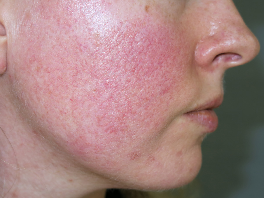
Erythema (from the Greek erythros, meaning red) is redness of the skin or mucous membranes, caused by hyperemia (increased blood flow) in superficial capillaries. It occurs with any skin injury, infection, or inflammation. Examples of erythema not associated with pathology include nervous blushes. Wikipedia
Erythema in rosacea usually involves the Butterfly or T-Zone area of the face.Image - Wikipedia Commons
Persistent Facial Erythema (PFE)
Dr. Del Rosso points out that if "...persistent facial erythema (PFE) is the pivotal diagnostic feature of cutaneous rosacea, including in both the presence or absence of papulopustular lesions..."that dermatologists"...Consider the role of PFE in essentially all patients with cutaneous rosacea." [1]Erythema used to be designated into one subtype along with telangiectasia: Erythematotelangiectatic Rosacea - Subtype 1 (now separated into Phenotype 2 and Phenotype 3). Read this notice about the subtype classification.
End Notes
[1] Journal of drugs in dermatology: JDD
June 2019 | Volume 18 | Issue 6 | Editorials | 503 | Copyright © June 2019
What Is “PFE”? It May Just Be Time You Found Out….
Del Rosso Q. James DO
JDR Dermatology Research/Thomas Dermatology Las Vegas, NV -
To understand why phenotype based treatment is superior to subtype based treatment for rosacea, lets take the case of classifying rosacea as Subtype 1 - Erythematotelangiectatic Rosacea.
A rosacean could present clinical features of erythema without any flushing symptoms and be treated with brimonodine. Another rosacean could present with clinical features of teleangiectasia and be treated with laser. These two very different clinical features are classified into one subtype.
A third rosacean could present with erythema due to flushing with no telangiectatic signs and treated with anti-flushing drugs.
The advantage of a phenotype based treatment is that these three clinical features are separated into (1) Phenotype Flushing, (2) Phenotype Persistent Erythema or (3) Phenotype Telangiectasia and receive totally different treatments determined by this new phenotype based treatment protocol.
"The characteristic rosacea symptoms manifest primarily, but not exclusively centrofacially, with forehead, nose, chin and cheeks significantly affected. Based on the various main symptoms a classification of the individual clinical pictures can be performed. However, a classification often does not reflect the clinical reality, since the various symptoms commonly coexist. The present review provides an introduction on pathogenesis and clinical manifestations of rosacea and prefers a symptom-oriented therapy approach." [1]
Subtype Classification Overlaps Signs and Symptoms
"However, a classification often does not reflect the clinical reality, since the various symptoms commonly coexist. The present review provides an introduction on pathogenesis and clinical manifestations of rosacea and prefers a symptom-oriented therapy approach." [2]
Since 2002, "A subtype-directed approach to treating rosacea patients is recommended to dermatologists." Wikipedia The subtype classification has proved wanting and was controversial from the beginning of its inception.
"Subtyping of rosacea, a post-hoc means of grouping more common presentations, can be and has been subverted inappropriately to imply strict categories without adequate consideration of the varying phenotypic presentation of individuals and the potential for temporal variation." [3]
Phenotype Classification Uses Signs and Symptoms Better
Phenotype Classification Distinguishes Signs and Symptoms
"The panel agreed on phenotype-based treatments for signs and symptoms presenting in individuals with rosacea." [4]
"It’s more important to be categorizing based on patients’ presentation in terms of signs and symptoms,” [5]
"The use of a phenotype approach will allow a more accurate and stringent classification of rosacea. New diagnostic tools emerge, and transcriptome analysis has revealed a possible distinct gene profile for each subtype of rosacea. A change in paradigm towards a phenotype classification, which can also easily be combined with newer diagnostic methods, such as transcriptome analysis, is an interesting possibility. The use of more objective criteria will allow those aspects that are most troubling to patients to be better targeted and allow the best treatment to be selected." [6]
"For past decade, a subtype classification has been used, but now rosacea experts are advising a move toward a phenotype approach which allows better targeting of treatment." [7]
What distinguishes the phenotype classification from the subtype classification? Answer.
Conclusion
Since 2016, a phenotype-directed approach to treating rosacea was introduced by the ROSCO panel which has proved superior and there is no controversy about this since it takes a symptom oriented approach. In December 2018 a group published in the British Journal of Dermatology concluded, "We present the most current evidence for rosacea management based on a phenotype-led approach." [8]
End Notes
[1] Pathogenesis and clinical presentation of rosacea as a key for a symptom-oriented therapy.
J Dtsch Dermatol Ges. 2016 Dec;14 Suppl 6:4-15
Reinholz M, Ruzicka T, Steinhoff M, Schaller M, Gieler U, Schöfer H, Homey B, Lehmann P, Luger TA[2] J Dtsch Dermatol Ges. 2016 Dec;14 Suppl 6:4-15
Pathogenesis and clinical presentation of rosacea as a key for a symptom-oriented therapy.
Reinholz M, Ruzicka T, Steinhoff M, Schaller M, Gieler U, Schöfer H, Homey B, Lehmann P, Luger TA"Most current guidelines are based on the identification of the rosacea subtype to select the appropriate therapy. However, in reality there is often an overlap of clinical features across rosacea subtypes in each patient, requiring several therapeutic strategies for optimal outcome. Thus, there is no single best way to treat all rosacea patients."
Dermatoendocrinol. 2017; 9(1): e1361574.
Rosacea: Epidemiology, pathogenesis, and treatment
Barbara M. Rainer, Sewon Kang, and Anna L. Chien[3] Shortcomings in rosacea diagnosis and classification
[4] Br J Dermatol. 2016 Nov 12. doi: 10.1111/bjd.15173.
Rosacea treatment update: Recommendations from the global ROSacea COnsensus (ROSCO) panel.
Schaller M, Almeida L, Bewley A, Cribier B, Dlova N, Kautz G, Mannis M, Oon H, Rajagopalan M, Steinhoff M, Thiboutot D, Troielli P, Webster G, Wu Y, van Zuuren E, Tan J.[5] Phenotype Classification Uses Signs and Symptoms Better
[6] Forum for Nord Derm Ven 2017, Vol. 22, No. 1
Rosacea: Time for a New Approach • Rosacea-Time-for-a-New-Approach.pdf
CARSTEN SAUER MIKKELSEN, PETER BJERRING, MARGARETA LIRVALL, MARGARETA SVENSSON, HELENE RINGE HOLMGREN, ALEXANDER SALAVA AND THEIS HULDT-NYSTRØM]7] J Drugs Dermatol. 2019 Sep 01;18(9):888-894
Recognizing Rosacea: Tips on Differential Diagnosis
Johnson SM, Berg A, Barr C[8] Br J Dermatol. 2018 Dec 26. doi: 10.1111/bjd.17590. [Epub ahead of print]
Interventions for rosacea based on the phenotype approach: an updated systematic review including GRADE assessments.
van Zuuren EJ, Fedorowicz Z, Tan J, van der Linden MMD, Arents BWM, Carter B, Charland L. -
Phenotype 3
Telangiectasias /tɛlˌæn.dʒiː.ɛkˈteɪ.zi.ə/ (sometimes called t. langiatasis) also known as spider veins or angioectasias, are small dilated blood vessels near the surface of the skin or mucous membranes, measuring between 0.5 and 1 millimeter in diameter.Telangiectasias was lumped together into one previous subtype along with erythema - Erythematotelangiectatic Rosacea - Subtype 1 (now separated into Phenotype 2 [Erythema] or Phenotype 3 [Telangiectasias]). Read this notice about the subtype classification.
Rosacea Telangiectasis courtesy of BPACThese dilated blood vessels can develop anywhere on the body but are commonly seen on the face around the nose, cheeks, and chin. Wikipedia
By Permission of Wikipedia CommonsNiamatu Cosmetic Facial Surgery has a photo gallery of nose and cheek spider veins (scroll down to see the photo gallery).
Treatment for Phenotype 3
Laser and Radio frequency (keep scrolling below)
Tranexamic acid (TXA) Intradermal Microinjections
Reply to this Topic
There is a reply to this topic button somewhere on the device you are reading this post. If you never heard about this topic and you learned about it here first, wouldn't it be a gracious act on your part to show your appreciation for this topic by registering with just your email address and show your appreciation with a post? And if registering is too much to ask, could you post your appreciation for this topic by finding the START NEW TOPIC button in our guest forum where you don't have to register? We know how many have viewed this topic because our forum software shows the number of views. However, most rosaceans don't engage or show their appreciation for our website and the RRDi would simply ask that you show your appreciation, please, simply by a post.
-
-
-
5 Amazing Remedies for Rosacea Skin Problems, DesiBlitz
-
'I LOOKED LIKE A TOMATO' Beauty blogger ditches gluten and dairy to combat embarrassing rosacea flare ups, The Sun
-
Treatment guidelines updated for patients with rosacea, November 21, 2016, Medical Press
Phenotype-based treatments recommended for rosacea, Health Day News, Clinical Advisor
Expert Panel Provides Recommendations for Rosacea Treatment, MPR
-
The ROSCO panel has changed the direction of classifying rosacea from subtypes to phenotypes.
What is the difference between a phenotype based treatment vs a subtype based treatment?
Genetics
We have to begin with Genotype which "is the part (DNA sequence) of the genetic makeup of a cell, and therefore of an organism or individual, which determines a specific characteristic (phenotype) of that cell/organism/individual. Genotype is one of three factors that determine phenotype, the other two being inherited epigenetic factors, and non-inherited environmental factors." Wikipedia
Genotype-phenotype distinction
"The genotype–phenotype distinction is drawn in genetics. "Genotype" is an organism's full hereditary information. "Phenotype" is an organism's actual observed properties, such as morphology, development, or behavior." Wikipedia
A phenotype (from Greek phainein, meaning "to show", and typos, meaning "type") is the composite of an organism's observable characteristics or traits, such as its morphology, development, biochemical or physiological properties, phenology, behavior, and products of behavior (such as a bird's nest). Wikipedia
Watch this short whiteboard animation on the difference between genotype and phenotype:
Phenotype and Subtype in Medicine
Phenotype
A phenotype in medicine is "the observable properties of an organism that are produced by the interaction of the genotype and the environment." Merriam Webster Medical Dictionary
Endotype
"An endotype is a subtype of a condition, which is defined by a distinct functional or pathobiological mechanism. This is distinct from a phenotype, which is any observable characteristic or trait of a disease, such as morphology, development, biochemical or physiological properties, or behavior, without any implication of a mechanism. It is envisaged that patients with a specific endotype present themselves within phenotypic clusters of diseases." Wikipedia
Disease Entity
"The main concept in nosology is the disease entity. Normally there are two ways to define a disease entity: Manifestational criteria and Causal criteria.
Manifestational criteria. They are a set of criteria based in signs, symptoms and laboratory findings that define a disease. They define the disease by its symptoms and medical findings.
Causal criteria are a causal chain of events that define the disease describing how it develops. They describe the disease by its etiology." WikipediaSubtype
A subtype in medicine is "a type that is subordinate to or included in another type <the blood group subtypes> <subtypes of a disease>." Merriam Webster Medical Dictionary
When the NRS 'expert' committee in 2002 began the classification of rosacea into subtypes, they did this based upon good motive. As one paper on this subject put it, "Broadly construed, disease subtyping is the task of identifying subpopulations of similar patients that can guide treatment decisions for a given individual. (When the subtypes have been established to be causally associated with the underlying mechanism, these are also called endotypes) Boland and colleagues describe the concept of a “vero-type” (the Latin word vero means “true”) to represent the true population of similar patients for treatment purposes. What constitutes these vero-types and how they should be discovered remains an open question. An active and growing body of work has explored different approaches for identifying homogeneous patient subgroups ranging from qualitative—based on clinical observations alone—to quantitative models that integrate mea- surements from diverse high-throughput biotechnologies." [1]
"Traditionally, disease subtyping research has been conducted as a by-product of clinical experience, wherein a clinician noticed the presence of patterns or groups of outlier patients and performed a more thorough (retrospective or prospective) study to confirm their existence." [1]
Subtype Related to Phenotype in Health Records
According to the author of the above referenced paper it appears that subtyping is related to 'Electronic Health Records and Phenotyping' and states, "Thus, much research has focused on developing new algorithms for annotating patient records with this information; in the literature, this task of extracting various clinical attributes for each individual is referred to as phenotyping....It is important to remember that phenotyping facilitates cleaner, but still broad, categories of disease....Thus, recent efforts have built on phenotyping work to move away from analysis of broad disease categories and instead provide more descriptive definitions of the disease over time."
Why is the phenotype classification superior to the subtype classification? Answer.
End Notes
[1] AI and Health, Daniel B. Neill, H.J. Heinz III College, Carnegie Mellon University
-
"I looked it up and thought I would try a new supplement (and very expensive, unfortunately) called Histame which is supposed to help those who have trouble eating histamine rich foods. Well, guess what? My skin is not only clear but much, much less puffy. My eyes are not bloodshot but white. I look so much healthier. For me, it was Histamine Intolerance all along. That is probably why my skin and my diet were so interrelated, and why taking a mast cell inhibitor like quercetin helped me. Anyway, I am now one happy camper. I still stick to a low histamine diet, which most docs who treat Histamine Intolerance seem to recommend, but my face looks great. My whole appearance has improved. If you seem to react to high histamine foods (tomatoes, smoked fish, aged cheese, etc.) you might want to give it a whirl. Histame is very expensive, though, but I don't have much choice for myself. My body really seems to be lacking in DAO, one of the enzymes that helps the body deal with histamine.
Regards, Queta"Histame 30 caps Dietary Supplement Food Intolerance Support. Clinically Shown To Degrade Histamine From Food Naturally helps discomfort from: Pizza, Tomatoes, Wine/Beer, Fish, Aged Cheeses, Bananas, Chocolate, More.. Supplement Facts Serving Size: 1 Capsule Servings per Container: 30 Amount per Serving Vitamin C 10 mg Calcium (as calcium carbonate) 100 mg Diamine Oxidase Enzyme 4000 HDU 0.3 mg (as porcine kidney protein concentrate pellets) Other Ingredients: Cellulose, water. HDU = Histamine Digesting Units Lactose-free Wheat-free Gluten-free Soy-free RECOMMENDED: For best results, take one (1) or two (2) capsules within 15 minutes of consuming products causing food intolerance.
The RRDi is an Amazon Affiliate. By clicking below on the link and purchasing the item, the RRDi receives a small fee. Thanks. Please post your experience using this product in this thread.
-
"The panel agreed on phenotype-based treatments for signs and symptoms presenting in individuals with rosacea." Dr. Martin Schaller serves on the ROSCO panel as well as the RRDi MAC.
Br J Dermatol. 2016 Nov 12. doi: 10.1111/bjd.15173.
Rosacea treatment update: Recommendations from the global ROSacea COnsensus (ROSCO) panel.
Schaller M, Almeida L, Bewley A, Cribier B, Dlova N, Kautz G, Mannis M, Oon H, Rajagopalan M, Steinhoff M, Thiboutot D, Troielli P, Webster G, Wu Y, van Zuuren E, Tan J. -
ORACEA is indicated for the treatment of only inflammatory lesions (papules and pustules) of rosacea in adult patients. No meaningful effect was demonstrated for generalized erythema (redness) of rosacea. Limitations of Use: This formulation of doxycycline has not been evaluated in the treatment or prevention of infections. ORACEA should not be used for treating bacterial infections, providing antibacterial prophylaxis, or reducing the numbers or eliminating microorganisms associated with any bacterial disease. To reduce the development of drug-resistant bacteria as well as to maintain the effectiveness of other antibacterial drugs, ORACEA should be used only as indicated." Source
-
Usually you read that Soolantra is for Phenotype 4, however, the report below shows that Soolantra may improve Phenotype 2 and Phenotype 3.
"Our series demonstrates better results with moderate to severe papulopustular rosacea as in the cases studied in trials. Nevertheless, we have also observed some response in mild cases of erythematotelangiectatic rosacea as well. Tolerance of ivermectin cream is high and produces quick results."
Dermatol Online J. 2016 Aug 15;22(8). pii: 13030/qt9ks1c48n.
Treatment of rosacea with topical ivermectin cream: a series of 34 cases.
Mendieta Eckert M1, Landa Gundin N. -

"Minocycline 100mg is non-inferior to doxycycline 40mg in efficacy over a 16- week treatment period. Furthermore, at follow up, RosaQoL scores and PaGA were statistically significantly more improved in the minocycline group than in the doxycycline group, and minocycline 100mg gives longer remission than doxycycline 40mg. In this study there was no significant difference in safety between these treatments, however, based on previous literature minocycline has a lower risk/benefit ratio than doxycycline. Minocycline 100mg may be a good alternative treatment for those patients who, for any reason, are unable or unwilling to take doxycycline 40mg."
Br J Dermatol. 2016 Oct 31. doi: 10.1111/bjd.15155.
DOMINO, doxycycline 40mg vs minocycline 100mg in the treatment of rosacea: a randomised, single blinded, non-inferiority trial, comparing efficacy and safety.
van der Linden MM, van Ratingen AR, van Rappard DC, Nieuwenburg SA, Spuls PI.Monocycline "is a broad-spectrum tetracycline antibiotic, and has a broader spectrum than the other members of the group....Minocycline is the most lipid-soluble of the tetracycline-class antibiotics, giving it the greatest penetration into the prostate and brain, but also the greatest amount of central nervous system (CNS)-related side effects, such as vertigo." Wikipedia
Orał Minocycline (minolira) has been approved for treating acne which is a timed released minocycline hydrochloride.
Oracea, a frequently prescribed timed released doxycycline (another tetracycline), by Galderma, is reportedly used by many rosacea sufferers to control their rosacea. It may be possible that minolira may be approved for rosacea.
Topical Minocycline
There have been a number of pharmaceutical companies in clinical trials with developing a monocycline topical which we have been following for some time now in this thread, and Zilxi has been announced as the front runner in this race.Minocycline-Induced Lupus
If you are not aware of minocycline-induced lupus, you are now informed.
-
The RRDi endorses the phenotype based treatment for rosacea proposed by the ROSCOE Panel in November 2016.
“Given the overlap of rosacea features across subtypes and the fact that no single treatment completely addresses all rosacea features, the current approach of diagnosing and treating rosacea by subtype may hinder individualised patient management. A new approach is needed to bring us closer to helping each and every rosacea patient receive the right treatment according to their signs and symptoms.” Jerry Tan, MD
“While the rosacea management landscape has advanced, the current subtype-based view of the disease can hinder progress by limiting the way we consider treatment options. These new ROSCO recommendations should help to make a positive impact on future treatment development and ultimately help improve the lives of people with rosacea through a symptom-led approach.” Esther J. van Zuuren, MD
Global ROSacea COnsensus (ROSCO) expert panel calls for new approach to diagnosis and treatment of rosacea to improve outcomes for patients, BusinessWire
Br J Dermatol. First published 2016 Oct 8. doi: 10.1111/bjd.15122. Full Text published 2017 Feb;176(2):465-471. doi: 10.1111/bjd.15173. Wiley
Updating the diagnosis, classification and assessment of rosacea: Recommendations from the global ROSacea COnsensus (ROSCO) panel.
Tan J, Almeida L, Bewley A, Cribier B, Dlova N, Gallo R, Kautz G, Mannis M, Oon H, Rajagopalan M, Steinhoff M, Thiboutot D, Troielli P, Webster G, Wu Y, van Zuuren E, Schaller M.For more info on this subject, read the articles, Classification Of Rosacea Remains Controversial • Shortcomings in rosacea diagnosis and classification • Who is the ROSCO Panel? • Phenotype Classification, How does it work?
Download: pdf
-
Souris24 (Marie) claims that she followed the liver and gallbladder flush discussed in this book and it improved her rosacea. This was in December 2015. Marie's last post was on August 15, 2016 where she states, "I believe that it made my rosacea better because it cleared some inflammation that was making my rosacea worse but the root cause of my rosacea is something else."
By clicking below on the link and purchasing the item, the RRDi receives a small fee. Thanks. Please post your experience using this product in this thread. -
The RRDi is pleased to announce that Greg K. Sakamoto, MD, has been appointed to serve on the RRDi and has graciously volunteered to assist us with his busy schedule. The RRDi appreciates Dr. Sakamoto's volunteer spirit.
-
Washing face with dandriff shampoo can combat rosacea, By Joe and Teresa Graedon, November 12, 2016, Houston Chronicle
-
Diet doesn't just effect acne or rosacea, it also effects depression.
Diet is associated with the risk of depression
UNIVERSITY OF EASTERN FINLANDRuusunen, Anu. Diet and depression. Publications of the University of Eastern Finland. Dissertations in Health Sciences 2013; no 185.
ISSN: 1798-5714
ISBN: 978-952-61-1201-5


#/media/File:The_Alcohol_Flushing_Response.png)
Demodex Density Count - What are the Numbers?
in Demodectic Rosacea
Posted
Demodex Density Counts Higher in Rosaceans
In many papers on rosacea, demodex density counts are higher than in the normal population. Below are reports about this subject, but the consensus is that five or more demodex mites per square centimeter is when there is an issue with demodectic rosacea. The normal count is 1 to 2 demodex mites per square centimeter for non rosacea subjects or in normal human subjects.
One report says, "Studies have found that people suffering from rosacea tend to have more Demodex mites. Instead of 1 or 2 per square centimetre of skin, the number rises to 10 to 20."
These Microscopic Mites Live on Your Face, by Lucy Jones, May 8, 2015, BBC Earth
"The mean mite count was 49.8 (range 2 to 158) in patients with rosacea and 10.8 (range up to 97) in control subjects (p < 0.001); the highest density of mites was found on the cheeks. A statistically significant increase in mites was found in all subgroups of rosacea, being most marked in those with steroid-induced rosacea." [1]
"The mean mite density was 2.03 mites/visual field in the rosacea group (range 0-5, SD = 1.2) and 0.16 mites/visual field (range 0-2, SD = 0.52) in the control group." [2]
"A mean density of 0.7 Demodex folliculorum/cm2 was found in controls, 98% of whom had less than five Demodex/cm2. When all clinical types of rosacea were considered collectively, the density of Demodex was significantly higher in patients with rosacea than in controls (mean = 10.8/cm2; P < 0.001). When the various clinical types of rosacea were considered separately, Demodex density was statistically significantly higher than in controls only in the PPR patients (mean = 12.8/cm2; P < 0.001)." [3]
"The results of our study revealed that DME [direct microscopic examination] is a more sensitive method for detecting Demodex than SSSB [Standardized skin surface biopsy], especially in patients with diffuse pattern and suspected rosacea type. Further research is needed to confirm this finding." [4]
"Every human being carries a colony of 1000 to 2000 Demodex mites." [5]
"More than five mites per cm2 are assumed a positive diagnosis of demodicosis. The validity of this optimal threshold is rather artificial and weakly evidence-based." [5]
"SSSB and DME were used to measure Demodex mites density (Dd). For SSSB, a standard area of 1 cm2 was drawn on a slide with a waterproof pen. A drop of cyanoacrylic adhesive was then placed on the other side of the slide and the adhesive-bearing surface was applied to the skin for one minute. After allowing the adhesive to dry, the slide was removed gently with surface skin, clarified with one to two drops of immersion oil, and covered with a cover slip. For DME, a 1 cm2 sized affected skin area was squeezed using a comedo extractor. The sample obtained was transferred to a 10% potassium hydroxide drop and covered with a cover slip. Samples obtained using both methods were studied under an optical microscope (×40, ×100)." [6]
"Patients with rosacea had significantly higher prevalence and degrees of Demodex mite infestation than did control patients." [7]
"The diagnosis of demodicosis was made when compatible clinical manifestations of demodicosis was combined with a high Dd (>5D/cm2) by SSSB, or DME." [6]
"High numbers of Demodex induced pro-inflammatory cytokine secretion whereas lower numbers did not. Demodex mites have the capacity to modulate the TLR signalling pathway of an immortalised human sebocyte line. Mites have the capacity to secrete bioactive molecules that affect the immune reactivity of sebocytes. Increasing mite numbers influenced IL8 secretion by these cells." [8]
"In the case of cutaneous demodicosis the presence of 5 or more Demodex on 1 cm2 will significantly increase the risk of cutaneous demodicosis." [9]
There is evidence that decreasing the mite count improves rosacea.
"The present study shows that PDL significantly reduced Dd in facial skin with one session." [10]
"To overcome this limitation we evaluated a new quantitative method of evaluating the viability of Demodex mites by using scattered light intensity (SLI)." [11]
Demodex Brevis Higher Count Than Demodex Folliculorum in Cylindrical Dandruff Patients
"Demodex infestation rates were significantly higher in the rosacea group than acne vulgaris and seborrheic dermatitis groups and controls (p=0.001; p=0.024; p=0.001, respectively)." [14]
"Demodex mites may be found on normal skin with a density of <5 mites/cm2. A diagnosis of demodicosis or Demodex infestation is considered when clinical signs/symptoms appear and when more than 5 mites/cm2 are present or when they penetrate into the dermis." [19]
In one paper it was shown that the "demodex density was 18.1 ± 10.7 (min: 0 - max: 48, Q1:12 - Q3:22) per cm2 before the pro-yellow laser treatment in the cases of "27 females (79.4%) and 7 males (20.6%)." [21]
"Some authors consider the density of > 5 mites per follicle as a pathogenic criterion." [22]
Increased Blood Glucose Level Correlates Increase in Demodex Density Count
Do the Numbers Matter?
The answer to that question according to one report [12], if you use a skin scraping with a light microscope, is no, which the report says, "The severity of the condition does not depend on the quantitative load of the mites in the scrape." Other reports say otherwise.
However when using a 'Confocal laser scanning in vivo microscopy', the answer is yes, which this same report concludes, "Confocal laser scanning in vivo microscopy is an effective diagnostic method to detect Demodex mites that does not require preliminary preparation for analysis and allows detecting Demodex mites at the level of the spiky epidermis layer, which is not accessible for scarification, to identify the species belonging to the size of Demodex mites (from 100 up to 200 μm - Demodex brevis, 200 to 400 μm – Demodex folliculorum)." [12]
"Comparing the results obtained by light microscopy and confocal laser scanning in vivo microscopy in patients with rosacea and healthy people, in more cases Demodex mites are detected by confocal laser scanning in vivo microscopy, whereas scrape in these patients were negative." [12]
"As a result of the study, we found that it is difficult to detect the mite by light microscopy of scrape per 1 cm2 of skin." [12]
For more information on how to quantify demodex density go to the subheading, Quantification and Methods for Demodex Density Counts in this article.
One study on comparing Standardized skin surface biopsy (SSSB) with Superficial Needle-Scraping (SNS) concluded that "that SNS is a simple and convenient method for assessing Demodex density of pustules in PPR and can be a useful alternative or addition to SSSB for evaluation of Demodex-associated facial papulopustular eruptions." [13]
"Statistics on Demodex prevalence in any host are likely to be underestimated, as a single sample may fail to find mites which may be present at other locations on the individual’s skin, or mite population may be below the threshold of detection by molecular methods, resulting in false negatives." [23]
Tools to Detect or Quantify Demodex Density Counts
Cellophane Tape Method [20]
CLSM [Confocal laser scanning in vivo microscopy] [16]
DME [direct microscopic examination]
Dermoscopy [17]
Microscope (simple)
RCM [reflectance confocal microscopy] [18]
Skin Scraping [potassium hydroxide preparation of skin scrapings]
SLI [scattered light intensity]
SSSB [standardized skin surface biopsy]
SNS [superficial needle-scraping]
Supereyes Macro Lens-Disposable Dermatoscope
Supereyes Smartphone Microscope Camera Adapter
Thumbnail-squeezing method
CLSM - did your dermatologist use this device to quantify your demodex density count? According to a Russian study [12], the CLSM in vivo method is the best method of quantifying demodex density counts which needs to be validated by comparing the other tools used. Another paper in Thailand states that SSSB has been 'considered to be the gold standard technique' but after careful investigation that the Skin Scraping technique is just as valid. [15] The paper by Karabay et al shows phots of using SSSB. [19]
See also, Methods for Quantifying Demodex Mites.
Reply to this Topic
There is a reply to this topic button somewhere on the device you are reading this post.
End Notes
[1] J Am Acad Dermatol. 1993 Mar;28(3):443-8.
The Demodex mite population in rosacea.
Bonnar E, Eustace P, Powell FC.
[2] J Eur Acad Dermatol Venereol. 2001 Sep;15(5):441-4.
Increased density of Demodex folliculorum and evidence of delayed hypersensitivity reaction in subjects with papulopustular rosacea.
Georgala S, Katoulis AC, Kylafis GD, Koumantaki-Mathioudaki E, Georgala C, Aroni K.
[3] Br J Dermatol. 1993 Jun;128(6):650-9.
Density of Demodex folliculorum in rosacea: a case-control study using standardized skin-surface biopsy.
Forton F, Seys B.
[4] Ann Dermatol. 2017 Apr;29(2):137-142
Demodex Mite Density Determinations by Standardized Skin Surface Biopsy and Direct Microscopic Examination and Their Relations with Clinical Types and Distribution Patterns.
Yun CH, Yun JH, Baek JO, Roh JY, Lee JR
[5] Iran J Parasitol. 2017 Jan-Mar; 12(1): 12–21.
PMCID: PMC5522688
Human Permanent Ectoparasites; Recent Advances on Biology and Clinical Significance of Demodex Mites: Narrative Review Article
Dorota LITWIN, WenChieh CHEN, Ewa DZIKA, and Joanna KORYCIŃSKA
Another paper concluded:
"Determination of five or more parasites in 1 cm2 area was considered as positive."
Medicina (Kaunas). 2020 Mar; 56(3): 107.
Pre-Treatment and Post-Treatment Demodex Densities in Patients under Immunosuppressive Treatments
Hacer Keles, Esra Pancar Yuksel, Fatma Aydin, and Nilgun Senturk
[6] Ann Dermatol. 2017 Apr; 29(2): 137–142.
Published online 2017 Mar 24. doi: 10.5021/ad.2017.29.2.137
PMCID: PMC5383737
Demodex Mite Density Determinations by Standardized Skin Surface Biopsy and Direct Microscopic Examination and Their Relations with Clinical Types and Distribution Patterns
Chul Hyun Yun, Jeong Hwan Yun, Jin Ok Baek, Joo Young Roh, and Jong Rok Lee
[7] "Twenty-three case-control studies included 1513 patients with rosacea. Compared with the control patients, patients with rosacea were more likely to be infested by Demodex mites [odds ratio, 9.039; 95% confidence interval (CI), 4.827-16.925] and had significantly higher Demodex density (SMD, 1.617; 95% CI, 1.090-2.145). Both erythematotelangiectatic rosacea (SMD, 2.686; 95% CI, 1.256-4.116) and papulopustular rosacea (SMD, 2.804; 95% CI, 1.464-4.145) had significantly higher Demodex density than did healthy control patients."
JAAD, September 2017 Volume 77, Issue 3, Pages 441–447.e6
Role of Demodex mite infestation in rosacea: A systematic review and meta-analysis
Yin-Shuo Chang, MD, Yu-Chen Huang, MD
[8] Demodex mites modulate sebocyte immune reaction: Possible role in the pathogenesis of rosacea.
Br J Dermatol. 2018 Mar 12;:
Lacey N, Russell-Hallinan A, Zouboulis CC, Powell FC
[9] Arch Med Sci. 2018 Mar; 14(2): 353–356.
Published online 2016 Jun 17. doi: 10.5114/aoms.2016.60663; PMCID: PMC5868666
The impact of age, sex, blepharitis, rosacea and rheumatoid arthritis on Demodex mite infection
Aleksandra Sędzikowska, Maciej Osęka, and Piotr Skopiński
[10] J Cosmet Laser Ther. 2018 Jun 08;:1-4
The rapid effect of pulsed dye laser on demodex density of facial skin.
Ertaş R, Yaman O, Akkuş MR, Özlü E, Avcı A, Ulaş Y, Ozyurt K, Atasoy M
[11] Exp Appl Acarol. 2019 Apr 19;:
Evaluation of Demodex mite viability using motility and scattered light intensity.
Gatault S, Foley R, Shiels L, Powell FC
[12] Dermatol Reports. 2019 Jan 23; 11(1): 7675.
Clinical picture, diagnosis and treatment of rosacea, complicated by Demodex mites
Alexey Kubanov, Yuliya Gallyamova, and Anzhela Kravchenko
[13] J Cosmet Dermatol. 2019 Jul 25. doi: 10.1111/jocd.13082. [Epub ahead of print]
A new superficial needle-scraping method for assessing Demodex density in papulopustular rosacea.
Huang HP, Hsu CK, Lee JY.
[14] An Bras Dermatol. 2020 Feb 12;:
Demodex folliculorum infestations in common facial dermatoses: acne vulgaris, rosacea, seborrheic dermatitis.
Aktaş Karabay E, Aksu Çerman A
[15] Indian J Dermatol Venereol Leprol. 2016 Sep-Oct;82(5):519-22. doi: 10.4103/0378-6323.174423.
Skin scrapings versus standardized skin surface biopsy to detect Demodex mites in patients with facial erythema of uncertain cause - a comparative study.
Bunyaratavej S, Rujitharanawong C, Kasemsarn P, Boonchai W, Muanprasert C, Matthapan L, Leeyaphan C.
[16] Br J Dermatol. 2012 Jun 20. doi: 10.1111/j.1365-2133.2012.11096.x.
Non-invasive in vivo detection and quantification of Demodex mites by confocal laser scanning microscopy.
Sattler EC, Maier T, Hoffmann VS, Hegyi J, Ruzicka T, Berking C.
What is confocal laser scanning microscopy?
[17] Dermatol Pract Concept. 2017 Jan; 7(1): 35–38.
Published online 2017 Jan 31. doi: 10.5826/dpc.0701a06
PMCID: PMC5315038
Usefulness of dermoscopy in the diagnosis and monitoring treatment of demodicidosis
Paula Friedman, Emilia Cohen Sabban, and Horacio Cabo
Int J Dermatol. 2010 Sep;49(9):1018-23.
Dermoscopy as a diagnostic tool in demodicidosis.
Segal R1, Mimouni D, Feuerman H, Pagovitz O, David M.
[18] RCM • Distinguishing rosacea from sensitive skin by reflectance confocal microscopy.
Skin Res Technol. 2014 Feb 13.
Reflectance confocal microscopy vs. standardized skin surface biopsy for measuring the density of Demodex mites.
Turgut Erdemir A, Gurel MS, Koku Aksu AE, Bilgin Karahalli F, Incel P, Kutlu Haytoğlu NS, Falay T.
What is reflectance confocal microscopy?
A paper on RCM states, “In its current form, RCM seems of limited value for noninvasive follow-up of rosacea inclinical practice.”
"RCM enables anti-inflammatory effect monitoring of topical ivermectin by determining mite presence. Quantifying exact mite number, and inflammatory and vascular characteristics is challenging due to device limitations. In its current form, RCM seems of limited value for noninvasive follow-up of rosacea in clinical practice."
J Dermatolog Treat. 2020 Mar 19;:1-9
Value of reflectance confocal microscopy for the monitoring of rosacea during treatment with topical ivermectin.
Logger JGM, Peppelman M, van Erp PEJ, de Jong EMGJ, Nguyen KP, Driessen RJB
[19] An Bras Dermatol. 2020 Mar-Apr; 95(2): 187–193.
Demodex folliculorum infestations in common facial dermatoses: acne vulgaris, rosacea, seborrheic dermatitis
Ezgi Aktaş Karabay and Aslı Aksu Çerman
[20] The cellophane tape method (placing tape on the face to stick to the mites)
Ubiquity and Diversity of Human-Associated Demodex Mites, paragraph 2
[21] The effect of 577-nm pro-yellow laser on demodex density in patients with rosacea
[22] Indian J Dermatol. 2014 Jan-Feb; 59(1): 60–66.
Human Demodex Mite: The Versatile Mite of Dermatological Importance
Parvaiz Anwar Rather and Iffat Hassan
[23] Journal of European Academy of Dermatology and Venereology [Full Text]
Demodex: a skin resident in man and his best friend
R. Foley, P. Kelly, S. Gatault, F. Powell